

Water Vascular System In Echinoderms
20.5: Echinoderms and Chordates
- Folio ID
- 65697
Deuterostomes include the phyla Echinodermata and Chordata (which includes the vertebrates) and two smaller phyla. Deuterostomes share similar patterns of early development.
Echinoderms
Echinodermata are named for their spiny pare (from the Greek "echinos" meaning "spiny" and "dermos" meaning "skin"). The phylum includes about 7,000 1 described living species, such as sea stars, ocean cucumbers, sea urchins, sand dollars, and brittle stars. Echinodermata are exclusively marine.
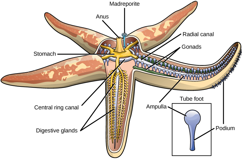
Adult echinoderms exhibit pentaradial symmetry and have a calcareous endoskeleton made of ossicles (Figure \(\PageIndex{1}\)), although the early larval stages of all echinoderms take bilateral symmetry. The endoskeleton is developed past epidermal cells, which may likewise possess pigment cells, giving vivid colors to these animals, likewise as cells laden with toxins. These animals accept a truthful coelom, a portion of which is modified into a unique circulatory system called a h2o vascular system. An interesting characteristic of these animals is their power to regenerate, fifty-fifty when over 75 per centum of their trunk mass is lost.
Physiological Processes of Echinoderms
Echinoderms have a unique system for gas exchange, food apportionment, and locomotion called the water vascular organisation. The arrangement consists of a central ring culvert and radial canals extending forth each arm. Water circulates through these structures allowing for gas, nutrient, and waste exchange. A structure on tiptop of the body, called the madreporite, regulates the amount of water in the h2o vascular organization. "Tube feet," which protrude through openings in the endoskeleton, may be expanded or contracted using the hydrostatic pressure in the arrangement. The arrangement allows for slow motion, just a great deal of power, as witnessed when the tube feet latch on to opposite halves of a bivalve mollusk, like a clam, and slowly, but surely pull the shells apart, exposing the mankind within.
The echinoderm nervous system has a nerve band at the center and five radial nerves extending outward along the arms. There is no centralized nervous control. Echinoderms have dissever sexes and release their gametes into the water where fertilization takes identify. Echinoderms may likewise reproduce asexually through regeneration from body parts.
Echinoderm Diversity
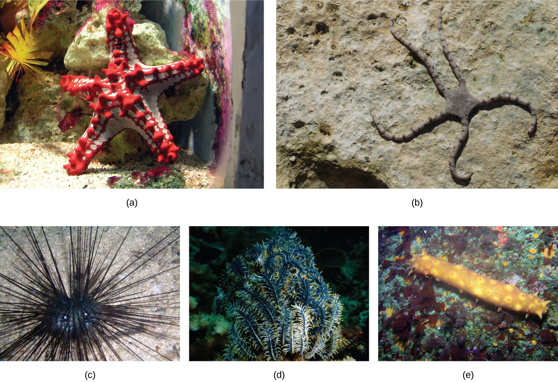
This phylum is divided into five classes: Asteroidea (sea stars), Ophiuroidea (brittle stars), Echinoidea (body of water urchins and sand dollars), Crinoidea (sea lilies or feather stars), and Holothuroidea (sea cucumbers) (Effigy \(\PageIndex{two}\)).
Maybe the best-known echinoderms are members of the class Asteroidea, or sea stars. They come up in a large variety of shapes, colors, and sizes, with more than ane,800 species known. The characteristics of sea stars that set them apart from other echinoderm classes include thick artillery that extend from a fundamental disk where organs penetrate into the artillery. Sea stars utilise their tube anxiety non just for gripping surfaces but besides for grasping prey. Sea stars have two stomachs, one of which they can evert through their mouths to secrete digestive juices into or onto prey before ingestion. This process can essentially liquefy the prey and make digestion easier.
CONCEPT IN ACTION
View this video to explore a sea star's body programme up close, lookout i move across the sea flooring, and run across it devour a mussel.
Brittle stars have long, thin arms that do not incorporate any organs. Sea urchins and sand dollars do not take arms only are hemispherical or flattened with five rows of tube feet, which assist them in slow movement. Sea lilies and plumage stars are stalked suspension feeders. Sea cucumbers are soft-bodied and elongate with five rows of tube feet and a series of tube feet around the oral cavity that are modified into tentacles used in feeding.
Chordates
The majority of species in the phylum Chordata are found in the subphylum Vertebrata, which include many species with which we are familiar. The vertebrates incorporate more than sixty,000 described species, divided into major groupings of the lampreys, fishes, amphibians, reptiles, birds, and mammals.
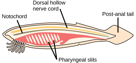
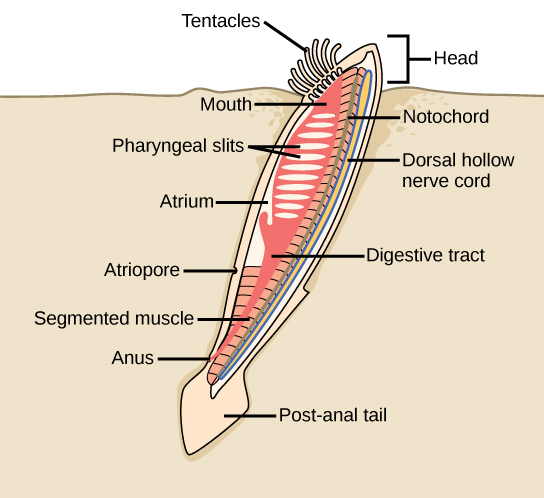
Animals in the phylum Chordata share iv key features that appear at some stage of their development: a notochord, a dorsal hollow nerve cord, pharyngeal slits, and a post-anal tail (Effigy \(\PageIndex{3}\)). In certain groups, some of these traits are nowadays only during embryonic development.
The chordates are named for the notochord, which is a flexible, rod-shaped structure that is establish in the embryonic stage of all chordates and in the adult stage of some chordate species. Information technology is located between the digestive tube and the nervus string, and provides skeletal back up through the length of the body. In some chordates, the notochord acts every bit the primary axial support of the body throughout the animal'southward lifetime. In vertebrates, the notochord is nowadays during embryonic development, at which fourth dimension it induces the development of the neural tube and serves as a back up for the developing embryonic body. The notochord, however, is not found in the postnatal stage of vertebrates; at this point, it has been replaced by the vertebral column (the spine).
The dorsal hollow nerve string is derived from ectoderm that sinks beneath the surface of the skin and rolls into a hollow tube during development. In chordates, it is located dorsally to the notochord. In contrast, other animal phyla possess solid nerve cords that are located either ventrally or laterally. The nervus cord found in about chordate embryos develops into the brain and spinal string, which compose the key nervous system.
Pharyngeal slits are openings in the pharynx, the region just posterior to the rima oris, that extend to the outside environs. In organisms that alive in aquatic environments, pharyngeal slits allow for the get out of water that enters the mouth during feeding. Some invertebrate chordates use the pharyngeal slits to filter food from the water that enters the oral cavity. In fishes, the pharyngeal slits are modified into gill supports, and in jawed fishes, jaw supports. In tetrapods, the slits are further modified into components of the ear and tonsils, since there is no longer whatsoever need for gill supports in these air-breathing animals. Tetrapod ways "four-footed," and this group includes amphibians, reptiles, birds, and mammals. (Birds are considered tetrapods because they evolved from tetrapod ancestors.)
The mail-anal tail is a posterior elongation of the body extending beyond the anus. The tail contains skeletal elements and muscles, which provide a source of locomotion in aquatic species, such as fishes. In some terrestrial vertebrates, the tail may also function in residuum, locomotion, courting, and signaling when danger is near. In many species, the tail is absent or reduced; for example, in apes, including humans, information technology is present in the embryo, merely reduced in size and nonfunctional in adults.
Fine art Connexion
Which of the following statements about common features of chordates is true?
- The dorsal hollow nervus cord is function of the chordate key nervous system.
- In vertebrate fishes, the pharyngeal slits become the gills.
- Humans are not chordates because humans do non have a tail.
- Vertebrates exercise non have a notochord at whatever point in their development; instead, they have a vertebral column.
Invertebrate Chordates
In addition to the vertebrates, the phylum Chordata contains two clades of invertebrates: Urochordata (tunicates) and Cephalochordata (lancelets). Members of these groups possess the 4 distinctive features of chordates at some betoken during their development.
The tunicates (Figure \(\PageIndex{4}\)) are also called sea squirts. The name tunicate derives from the cellulose-like carbohydrate fabric, called the tunic, which covers the outer body. Although tunicates are classified equally chordates, the developed forms are much modified in body plan and do non accept a notochord, a dorsal hollow nerve string, or a mail-anal tail, although they do have pharyngeal slits. The larval class possesses all four structures. Most tunicates are hermaphrodites. Tunicate larvae hatch from eggs within the developed tunicate's torso. Later hatching, a tunicate larva swims for a few days until it finds a suitable surface on which it tin can attach, usually in a dark or shaded location. Information technology then attaches by the head to the substrate and undergoes metamorphosis into the developed form, at which signal the notochord, nerve string, and tail disappear.
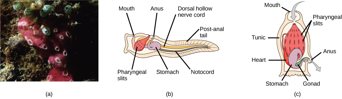
Most tunicates live a sessile existence in shallow sea waters and are intermission feeders. The primary foods of tunicates are plankton and detritus. Seawater enters the tunicate'southward body through its incurrent siphon. Suspended cloth is filtered out of this water past a mucus net (pharyngeal slits) and is passed into the intestine through the action of cilia. The anus empties into the excurrent siphon, which expels wastes and h2o.
Lancelets possess a notochord, dorsal hollow nervus string, pharyngeal slits, and a post-anal tail in the adult stage (Figure \(\PageIndex{5}\)). The notochord extends into the caput, which gives the subphylum its proper noun (Cephalochordata). Extinct fossils of this subphylum engagement to the centre of the Cambrian flow (540–488 mya).The living forms, the lancelets, are named for their blade-like shape. Lancelets are merely a few centimeters long and are usually found buried in sand at the bottom of warm temperate and tropical seas. Similar tunicates, they are suspension feeders.
Section Summary
Echinoderms are deuterostome marine organisms. This phylum of animals acquit a calcareous endoskeleton equanimous of ossicles covered past a spiny peel. Echinoderms possess a water-based circulatory system. The madreporite is the indicate of entry and exit for water for the water vascular arrangement.
The characteristic features of Chordata are a notochord, a dorsal hollow nerve string, pharyngeal slits, and a post-anal tail. Chordata contains two clades of invertebrates: Urochordata (tunicates) and Cephalochordata (lancelets), together with the vertebrates. Nigh tunicates live on the ocean floor and are break feeders. Lancelets are pause feeders that feed on phytoplankton and other microorganisms.
Art Connections
Figure \(\PageIndex{3}\): Which of the following statements about common features of chordates is true?
A. The dorsal hollow nerve cord is part of the chordate central nervous arrangement.
B. In vertebrate fishes, the pharyngeal slits become the gills.
C. Humans are not chordates because humans practice not have a tail.
D. Vertebrates practise non have a notochord at any bespeak in their development; instead, they have a vertebral cavalcade.
- Respond
-
A
Glossary
- Cephalochordata
- a chordate clade whose members possess a notochord, dorsal hollow nervus string, pharyngeal slits, and a post-anal tail in the adult stage
- Chordata
- a phylum of animals distinguished by their possession of a notochord, a dorsal hollow nerve string, pharyngeal slits, and a post-anal tail at some bespeak during their development
- dorsal hollow nervus cord
- a hollow, tubular structure derived from ectoderm, which is located dorsal to the notochord in chordates
- Echinodermata
- a phylum of deuterostomes with spiny skin; exclusively marine organisms
- lancelet
- a member of Cephalochordata; named for its bract-similar shape
- madreporite
- a pore for regulating entry and exit of water into the water vascular organisation
- notochord
- a flexible, rod-shaped structure that is establish in the embryonic stage of all chordates and in the adult stage of some chordates
- pharyngeal slit
- an opening in the pharynx
- mail service-anal tail
- a muscular, posterior elongation of the body extending across the anus in chordates
- tetrapod
- a four-footed animal; includes amphibians, reptiles, birds, and mammals
- tunicate
- a sessile chordate that is a member of Urochordata
- Urochordata
- the clade equanimous of the tunicates
- vertebral column
- a series of dissever bones that surround the spinal cord in vertebrates
- h2o vascular system
- a organisation in echinoderms in which water is the circulatory fluid
Contributors and Attributions
-
Samantha Fowler (Clayton State University), Rebecca Roush (Sandhills Community College), James Wise (Hampton University). Original content by OpenStax (CC BY 4.0; Access for gratuitous at https://cnx.org/contents/b3c1e1d2-83...4-e119a8aafbdd).

Water Vascular System In Echinoderms,
Source: https://bio.libretexts.org/Courses/Folsom_Lake_College/BIOL_310%3A_General_Biology_(Wada)/zz%3A_Back_Matter/21%3A_BIOL_307_Modules/20%3A_Animal_Diversity/20.05%3A_Echinoderms_and_Chordates
Posted by: godwinaces1963.blogspot.com
0 Response to "Water Vascular System In Echinoderms"
Post a Comment